
Dentures, Full Set of False Teeth with Hollywood Bleach Shade Teeth Plutusdental Denture
Dentists use computed tomography (CT) scans to capture 3D dental X-rays of your teeth, jaws, joints, nerves and sinuses. These X-rays can also detect tumors or facial fractures. Surgeons often use dental CT scans to check the height, width and location of your jawbone before dental implant placement. Advertisement.
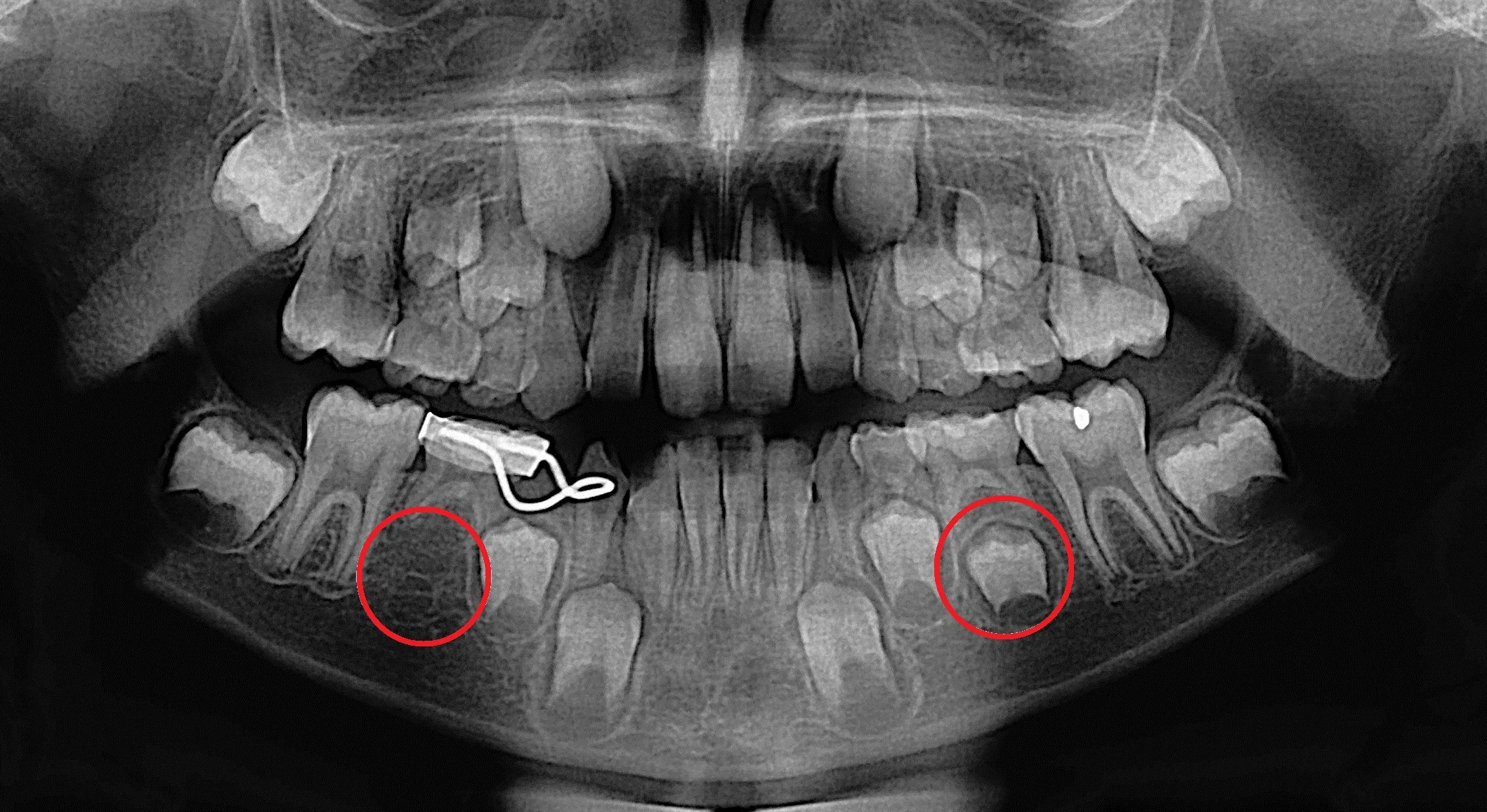
My impacted wisdom tooth Xray. r/mildlyinteresting
They are made by a panoramic X-ray machine that rotates around your head. The panoramic X-ray is commonly used to detect wisdom teeth; acquire more information about jaw issues and missing or extra teeth; help identify abscesses, cysts, and tumors; and evaluate growth and development in children. This type of X-ray also provides information.

FileXray chest cancer.jpg
Your exposure to radiation during a dental X-ray is extremely low. Dental X-rays emit a type of radiation that helps capture detailed images of your teeth and bones, which are crucial for diagnosis and treatment planning. The level of radiation is measured in millirems (mrem), and a typical dental X-ray gives off approximately 0.5 mrem per image.
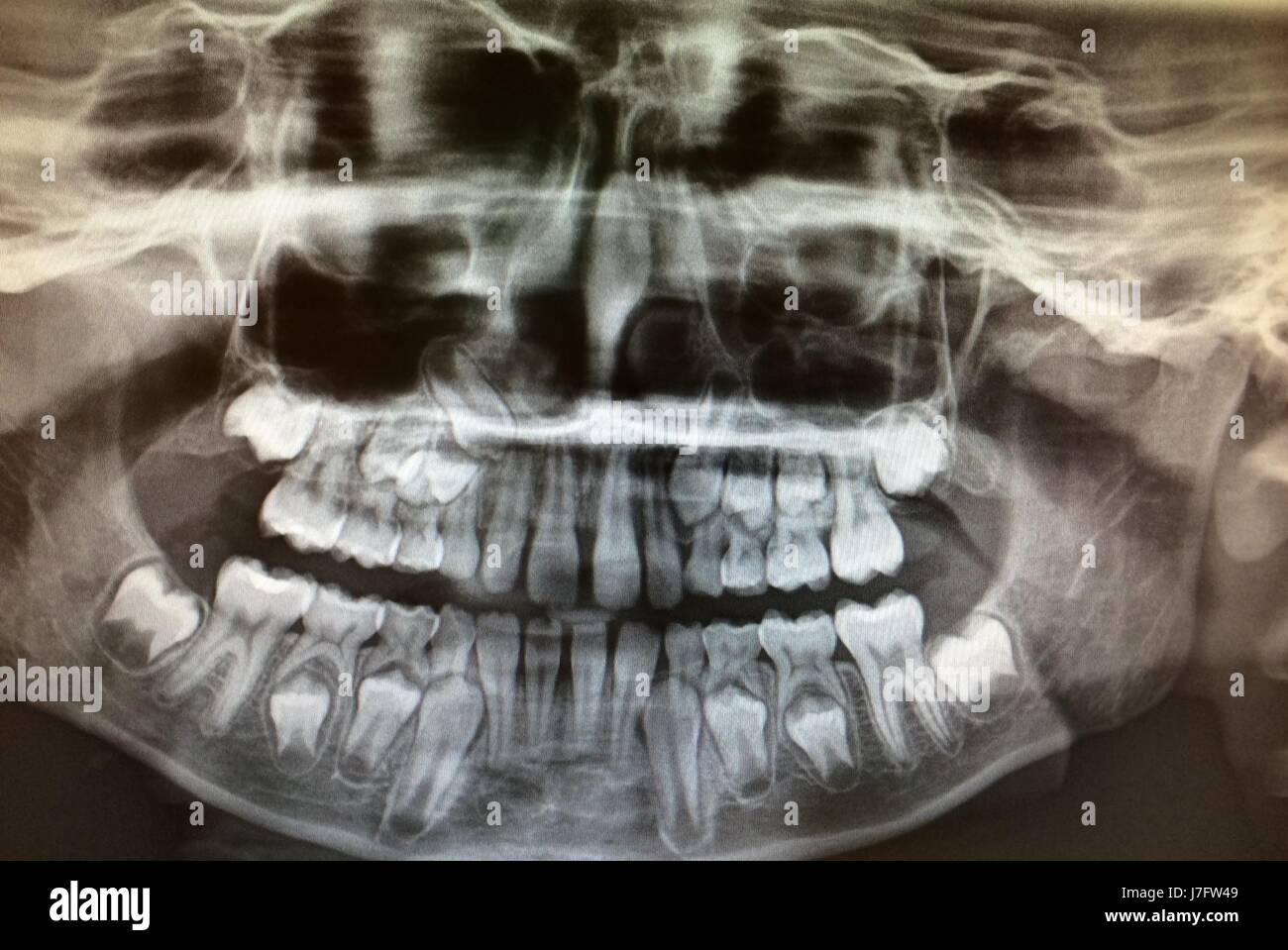
Panoramic Dental XRay of Childs Teeth Development Stock Photo Alamy
Dental radiographs, or X-rays, contain images of your teeth that a dentist can use to monitor your oral health. Low radiation levels create a still image of a patient's gums and teeth. X-rays allow dentists to identify problems or potential issues, such as cavities, impacted teeth, tooth decay, and more. X-rays may also determine if there's.

Xrays The Tooth Photos
3. Have the Patient Tilt Their Chin Up or Down, or Look Forward. Since the angulation of your tubehead needs to be extreme at times, you're going to need your patients to help. Not by holding the X-ray sensor, but by changing the position of their mouth. For example, if you need some lower PAs, have your patient tilt their chin way up in the air.
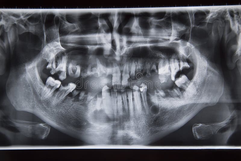
Dental xray with bad teeth stock image. Image of full 52812955
How to take a good dental x-ray is not only about proper technique. but actually understanding what you are looking for in the image is super important too.

How to Get Perfect Teeth
Also called radiographs, dental X-rays are images of your teeth that allow a dentist to assess your oral health. X-rays use low levels of radiation to capture photos of the inside of teeth and gums. This can help a dentist identify potential problems such as tooth decay, cavities and impacted teeth. Without X-rays, a dentist cannot identify.

Perfect teeth mirroring stock image. Image of reflection 53500833
X-rays should be taken to check for development of wisdom teeth. Adults with teeth. A full series of X-rays is indicated when there is evidence of dental disease or history of extensive decay. X.
xray image of a jaw with teeth Stock Photo Alamy
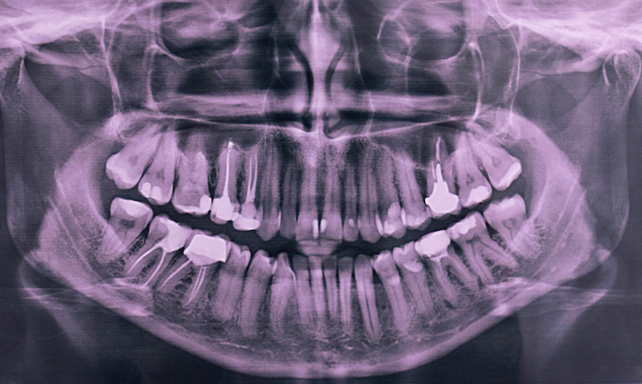
What is Panoramic X-ray? Panoramic radiography, also called panoramic x-ray, is a two-dimensional (2-D) dental x-ray examination that captures the entire mouth in a single image, including the teeth, upper and lower jaws, surrounding structures and tissues.. The jaw is a curved structure similar to that of a horseshoe. However, the panoramic x-ray produces a flat image of the curved structure.

Perfect Teeth (Album Mix) YouTube
Panoramic: large single x-ray that shows all the teeth and the jaw bone. This kind of x-ray is used a lot in orthodontics, and in evaluating missing teeth, extra teeth and wisdom teeth. Cone Beam CT: large single x-ray that shows a 3D image of the desired area. These CT scans do produce a little more radiation than the x-rays.
Changing smiles are normal and beautiful! — Lakes Region Dental Center
Here are a few of the most common types of X-rays carried out: Periapical Offers a view of the entire tooth, from the crown to the bone that helps to support the tooth.; Bite-Wing Provides a visual of both the lower and upper posterior teeth. This kind of X-ray shows the dental practitioner how these teeth touch one another (or occlude) and helps to figure out if decay is present in between.
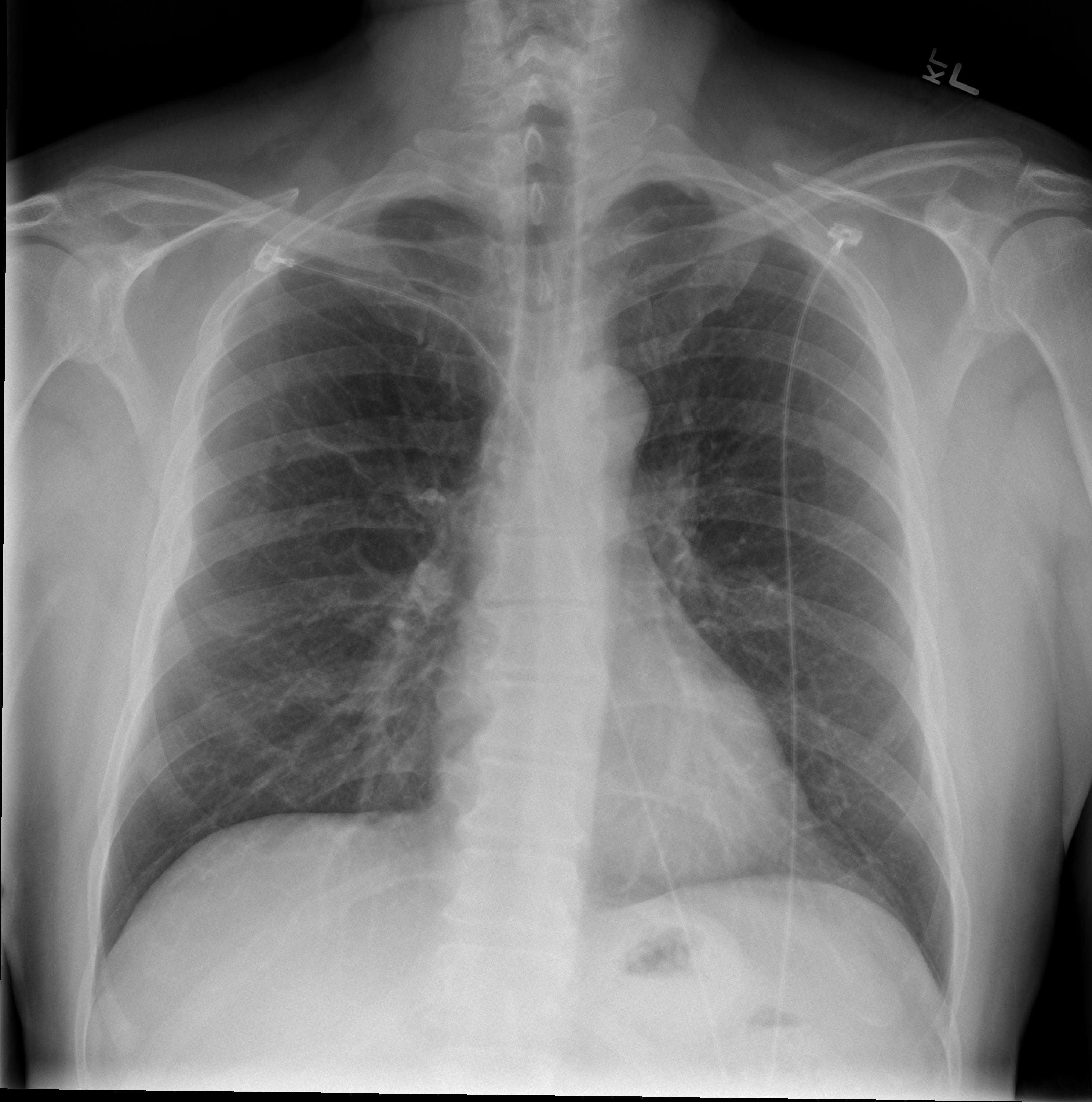
FileChest xray posteroanterior view.jpg Wikimedia Commons
Some of the things your dentist will examine in your dental X-rays include: Position, size, and number of teeth. Changes in the root canal. Bone loss in the jaw or facial bones. Bone fractures. Tooth decay, including between teeth or under fillings. Abscesses and cysts.
Are Dental XRays Safe?
Dental x-rays are used to make quick and painless images of your teeth and jaws. X-rays are invisible beams of energy, a form of radiation. The images are displayed on film or on the computer monitor (digital imaging) after the x-rays pass through an area of the body and are absorbed differently depending on the density of the structures.
Dental XRays Safety, Risks and Procedure Hove Dental Clinic
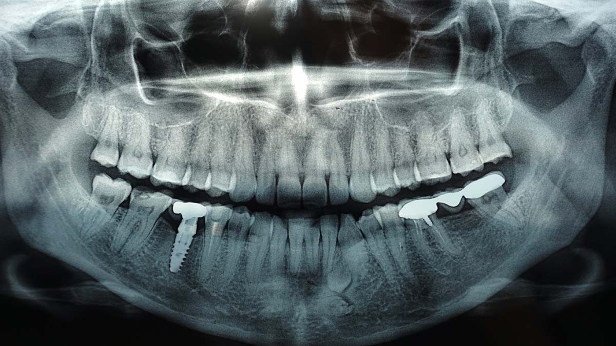
A panoramic dental x-ray captures a single image that shows your teeth, jawbones and surrounding facial structures. Dentists and oral surgeons use these x-rays to diagnose dental problems and plan treatments, especially for restorative dentistry like dental implants, or teeth straightening and orthodontic work.. Here you can discover how panoramic radiography works, what to expect from the.

Dental Xrays and Brain Tumors — Oh My! ScienceBased Medicine
Taking X-rays of diagnostic quality is easy to achieve — if the clinician follows basic, simple rules. Taking X-rays that are not in accord with the standards of dental practice may be considered negligence. Radiographs, or X-rays, are an integral part of dental practice. When radiographs are not of diagnostic quality, it can result in a.

What are the different types of Digital Dental Xrays? Palms Dentist, Shirley Christchurch
A dental X-ray is a type of radiograph used to capture images of the teeth, gums, and bones in the mouth. It is a valuable tool that allows dental professionals to diagnose and treat various dental conditions. When you have a dental X-ray, a small sensor or film is placed in your mouth, and an X-ray machine is used to take pictures of your teeth.